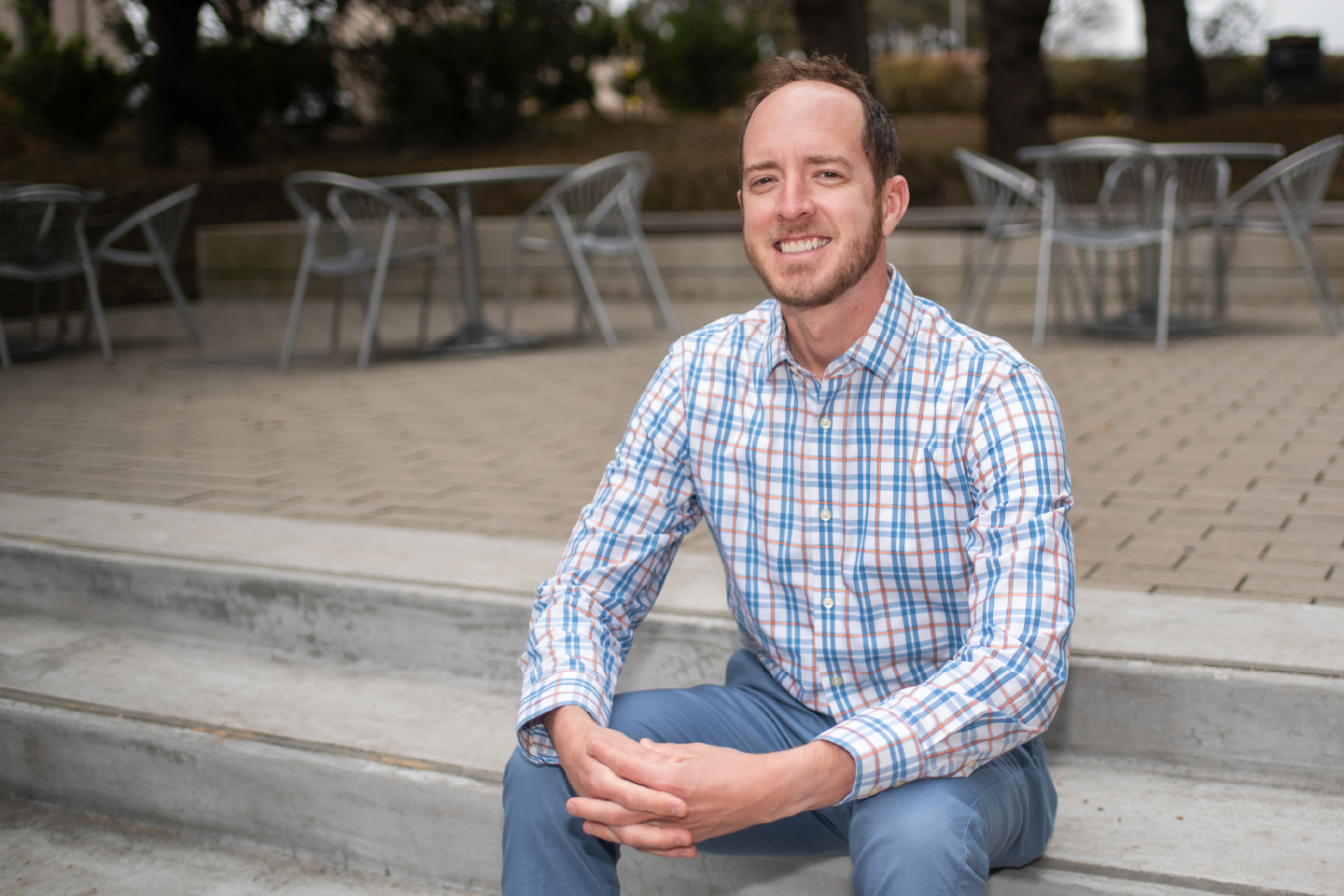
This news feature is part of Dell Med’s Voices, a series of profiles that highlight the people of Dell Med as they work to improve health with a unique focus on our community.

When looking at the results of an X-ray or MRI, Jack Virostko, Ph.D., sees more than just a “pretty” picture: He uncovers the story the image tells, picking apart the underlying medical data that can inform lifesaving decisions.
Virostko, assistant professor in Dell Medical School’s Department of Diagnostic Medicine and courtesy assistant professor in the Department of Oncology, transitioned from studying engineering to medical technology. He applies engineering fundamentals to diagnostic imaging so medical professionals can effectively treat conditions like cancer and diabetes.
From Bench to Bedside, & Back Again
After obtaining undergraduate and graduate degrees in engineering, Virostko didn’t find the inspiration he was looking for in bridges and engines — instead, he wanted to directly help patients. A Master of Science in Clinical Investigation provided formal training he needed in biostatistics, epidemiology and clinical trial design.
“In this program, I was the only Ph.D. scientist in classes full of M.D.s, which gave me a different perspective into my own work and medical research in general,” Virostko says.
His research career began in the lab, analyzing diabetes in mice. When he moved on to bringing medical technologies to human patients, Virostko suddenly had to consider factors like side effects and individual responses to therapy. This movement from basic research to clinical practice is called translational research.
“It’s often called ‘bench-to-bedside,” which is a nice catchy phrase that makes it sound deceptively simple,” Virostko says. “People are often trained in just the lab or just the human side of research, but it’s important to address both. My holistic training gives me insight into the challenges of getting technology from lab models all the way to human patients.”
Pathways to Personalized Medicine
Virostko’s research focuses on quantitative imaging, or the underlying data in a picture. For instance, when looking at the MRI of a tumor, he can look at the cell density that the instruments measure to tell if cells are dying at the site, indicating whether or not treatment is working. For Virostko, it’s about going beyond what’s visible to the eye.
“It’s the difference between stepping outside and saying ‘it’s cold today’ and reading a thermometer that tells you it’s 33 degrees outside,” Virostko says. “The measurements we can make with quantitative imaging help us form valuable benchmarks.”
What makes his work even more complex is the mystery surrounding cutting-edge technology. From the time a patient goes into the MRI scanner to the moment an image pops up on a technician’s screen, there are many steps in between. And researchers aren’t always privy to these interrelated steps, which are sometimes trade secrets.
“It’s a bit of a black box,” Virostko says. “When we adjust for one parameter — like the water content of a tumor— it changes the way we’re able to collect data from another part of the image, and we don’t always understand why. Imaging technology is a complex web.”
In one Dell Med research project, Virostko and his teammates are conducting MRI scans of breast cancer tumors as patients undergo therapy. Measurements allow teams to detect whether a tumor is responding to therapy and alter individual treatment plans based on a patient’s scan. Virostko is doing similar work with Type 1 diabetes, looking at the distribution of quantitative values to understand the way the pancreas changes throughout treatment.
“I think that in the future, getting an MRI of your whole body to screen for disease will be part of a routine annual exam,” Virostko says. “We are moving along the path to personalized medicine.”