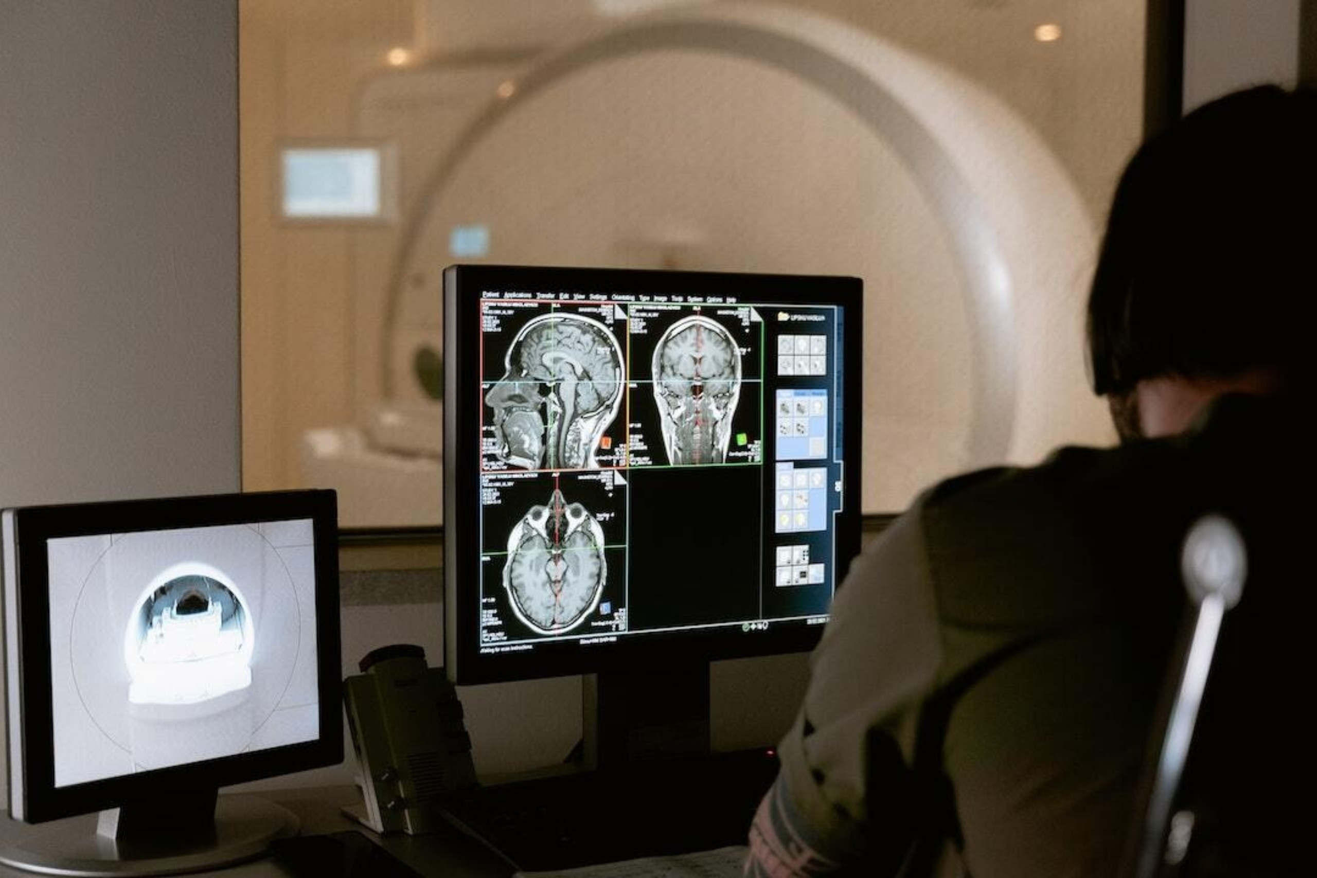
Diagnostic imaging is undergoing a transformation with the advent of artificial intelligence: Soon, patients can anticipate faster, more precise test results through tools that help doctors see more than meets the (human) eye.

Diagnostic radiology residents at Dell Medical School are taking advantage of built-in time and resources to advance AI in medical imaging.
Leading the way in Central Texas are physicians training within Dell Medical School’s Diagnostic Radiology Residency. As health and AI continue to converge, Dell Med’s radiology residents are engaging in research that assesses how — and how well — AI can empower radiology care.
“We’re at a time in which radiology is uniquely suited for the application of AI. It’s a field inherently based on large, complex sets of digital data that require lots of analysis to get diagnostic information out of,” says Jack Virostko, Ph.D., who directs research within the residency program and is an associate professor of biomedical engineering and diagnostic medicine at UT. “UT has a really strong AI infrastructure that includes National Science Foundation-supported institutes and faculty deeply interested in machine learning.
“In addition to typical didactic research training, our program helps put residents into research projects that leverage campus strengths and span disciplines but connect directly to diagnostic medicine. For the field of AI, it means helping residents research ways to implement these emerging tools, with impact for both patients and the practice of medicine.”
Faculty leaders like Virostko, all working to define the future of health, shape the program to afford trainees dedicated research time to explore the field’s digital- and data-driven nature, and learn where AI can benefit patients seeking diagnostics.
Testing & Validating Artificial Intelligence
As of May, the Food and Drug Administration has authorized the use of nearly 900 AI/machine learning-enabled medical devices, with most of them intended for radiology. Radiologists today are beginning to use these tools, which are trained on thousands of images, to more efficiently analyze CT scans, MRIs, tissue slides and more, giving faster diagnostic care to patients.
Looking to apply a more watchful eye, Christopher Kaufmann, M.D., is testing, benchmarking and validating the specialized information offered by large language models, a type of AI program that can interpret and generate data using natural language.
“Large language models rapidly evolve, and we want to keep pace with that,” says Kaufmann, a second-year radiology resident. “As AI becomes more ingrained in our lives, how can we trust it? Is the information it offers validated? That’s what we’re looking at now.”
For Kaufmann, constantly monitoring the performance of these tools is key if they are to be used in clinical settings, as diagnostic errors can have life-changing consequences.
Kaufmann demonstrated the powerful knowledge base that large language models have as well as the discrepancies within them: He presented a study at the Radiological Society of North America’s annual meeting showing that ChatGPT 4.0 scored over 90% correct on a series of practice questions from the American College of Radiology Diagnostic In-Training Exam. Other large language models such as Google Bard and Microsoft’s BingChat, as well as older versions of ChatGPT, scored lower in the 60% to 70% range.
Now, having launched his research through dedicated time for scholarly activity during residency training, Kaufmann aims to develop standardized tools so that any time a large language model evolves, it can be measured against human-based judgment in the field of radiology.
“Medicine is comprised of very specialized fields with lots of domain-specific knowledge. I’m interested in how these large language models perform the narrower you go, when you get to real-world questions and the subspecialty areas of medicine,” Kaufmann says. “There’s lots of interest in large language models, but before putting them to practice, you have to the address trust, credibility and reliability of their knowledge.”
Predicting Disease Through Incidental Data
Radiologists often discover more than they’re looking for: Imagine a patient undergoing imaging following a car accident, but while analyzing their back injury, the radiologist happens to see that the patient also has gallstones. These “incidental findings” — medical findings unrelated to the original purpose for imaging — are commonplace in diagnostics and continue to increase with advancements in technology.
Jack Webb, M.D., a first-year radiology resident, looks to apply the power of large language models toward incidental findings like gallstones to determine whether AI can predict the likelihood of developing related diseases — giving patients a better understanding of their health, and helping radiologists deliver higher-value care.
Webb’s research leverages AI’s ability to analyze images in far greater detail than the human eye. For patients with gallstones, looking deeper into the data may mean treating related conditions like cholecystitis before they ever become an emergency.
“Gallstones are a very common and expensive problem, and I wondered how many patients with incidental gallstones go on to have disease from them. My research looks at the characteristics of gallstones and the gallbladder with AI and uses algorithms to see if there’s any correlation between these characteristics and subsequent disease,” Webb says. “Radiologists could use this data to gain real-time information on risk factors, predict a patient’s chance of developing cholecystitis and better map their diagnostic care.
“With a radiologist who uses AI, patients could proactively address their incidental gallstones, which would be far more affordable and convenient for them than dealing with an unexpected, emergent case down the road.”